"Creating Beautiful Smiles"
How itero intraoral digital scanning technology offers major advantages for practitioners and patients
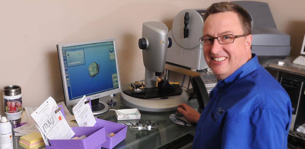
As digital technology advances, it offers a variety of new methods for dentists who seek to provide better experiences and more satisfactory outcomes for their patients. One technology that deserves considerably more attention from quality-seeking dental professionals is the internal oral digital scanner, such as the Itero family of Element™ Intraoral Scanners.
Today’s high-quality intraoral scanner systems simplify and speed the task of accurately modeling a patient’s oral cavity and utilizing this model to aid in performing restorative work. Intraoral digital scans provide highly accurate dental measurements in the form of secure, compact digital information files. These digital files can then be quickly transmitted to suitably-equipped laboratories, which can utilize them to fabricate prescription appliances much faster, easier, better, and cheaper than ever before.
This modern digital technology is easily applicable to crowns, veneers, bridgework, and implants. Orthodontists can also use today’s updated scanning technology to diagnose dental problems and provide the best possible treatments.
What’s more, from the patient’s perspective, the ease and comfort involved with using the new generation of intraoral scanning devices provide major improvements over traditional, relatively time-consuming, cumbersome, and messy oral modeling technologies.
In the past few years, a surprising number of dentists have converted their practices to these new dental scanners, and already have successfully recorded millions of dental scans with remarkable results that are much appreciated by their patients.
Dental Scanners
Today’s dental scanners are centered around a toothbrush-sized probe that the dental professional carefully runs along the surfaces of a patient’s teeth in order to record with remarkable precision their size, shape, and exact placement. These “wands” are extremely light in weight, comfortably shaped, and equipped with important and useful features like an anti-fog lens, auto-focus, zoom, freeze-frame, auto power-off, and super-wide-angle viewing.
Some of these systems are stand-alone and self-contained, while others are designed and built to connect with a separate – perhaps already existing – computer of the dentist’s choice.
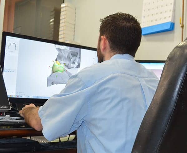
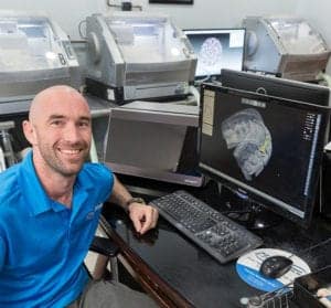
As the practitioner guides the scanning wand, the system displays whatever it is seeing on a conventional TV screen or computer monitor in real time. It also records the digital data for later review, study, sharing with colleagues and staff, and eventual transmission to any suitably-equipped fabrication facility.
Allowing the dental practitioner to view the digital images at the moment they are being captured provides a simple, intuitive opportunity to verify that the scan is both accurate and complete. This compares favorably with older methods of modeling teeth and gums that required having to wait several weeks before hearing back from the lab about any problems, glitches, or inaccuracies in the mold-making process.
The best systems, like those from Itero, no longer even require patients to powder their teeth with titanium dioxide.
Dental Scan Machine
The equipment required to perform these state-of-the-art oral scans consists of a hand-held scanning wand that houses a miniature imaging system. This wand is connected to a hardware module that both powers the scanner and receives the digital imagery being generated.
The scanner-bearing wand is truly the “business end” of the system, needing to be acceptably shaped and relatively unobtrusive for the patient’s comfort, as well as lightweight and a good fit for the hands of the professional using it.
Digital Scan
The scanning action is important, but so is the STL file produced by the scanning equipment. It is this file which the fabricating laboratory uses to actually build the required dental appliance.
Depending upon the laboratory’s equipment, the digital file can be used to drive computer-controlled milling machines to create the required appliances, or to control 3-D printers capable of extruding dental-quality material. Milling tends to be more accurate, but a precision 3-D printer can also be a good option for dental fabrication.
Digital Dental Impressions
Capturing a digital dental impression is not difficult, but does take a bit of practice.
One common approach is to handle the intraoral scanner as if it were a toothbrush. The biggest difference between a scanning wand and an actual toothbrush – aside from the absence of bristles – is that the operator should hold the scanning element as though it were a pen or pencil, from beneath. This is a natural, relaxed posture that permits extremely accurate placement and movement of the scanning device.
The operator holds the imaging portion of the digital scanning device flat against the surface of the teeth, touching gently as much as possible, and begins scanning at the occlusal surface of the most distal tooth. The idea is to gently move the scanning head toward the canine, then around toward the other canine, and then straight back into the mouth again towards the most distal tooth on the other side.
Retraction is made easier by the shape of the scanner itself, which ideally has been designed specifically to aid in this movement without pinching the lip between the scanner and the anterior teeth.
Another series of movements of the scanner then captures the digital data from the mesial and distal on the lingual surfaces, with the scanner manipulated slowly within the patient’s mouth, again keeping the scanning surface flush with the lingual.
After half a dozen or so practice passes on the mouths of cooperating assistants, most practitioners can become adept enough to quickly and easily complete accurate scans of an actual patient’s mouth.
Continue Reading…
- Everything you need to know about Valplast Dentures
- At-Home Teeth Whitening Products
How Does An Intraoral Scanner Work
Digital dental scanners were initially introduced in the 1980s, and have come a long way since then.
The newest intraoral scanners consist of a small, smooth wand attach to a computer running specialized software that captures the digital data sensed by the wand. The smaller the wand, the more easily it can reach all parts of the oral cavity and, in general, the more accurate and detailed the data it can capture. Not incidentally, smaller wands are more comfortable for patients, so they are less likely to induce a gag response or to require a patient to hold their mouth wide open during the scanning procedure.
The dental practitioner carefully inserts the wand into the patient’s mouth and glides it gently along the surfaces of the teeth. The wand captures data points as often as six thousand times each second, automatically registering the sizes and shapes of each tooth. It continuously sends this data to the connected computer’s software, which builds it into a complete and extremely precise impression of the patient’s oral cavity.
In just a minute or two of scanning, the system is capable of producing a detailed digital map that can be viewed on any suitably equipped computer and transmitted to a cooperating laboratory for use in fabricating any needed appliance.
What Are the Patient Advantages
Dentists who convert to intraoral digital scanning technology offer their patients significant advantages over those who still rely on outmoded methods of physically taking molds of the teeth.
These advantages include major savings of time and money, along with a much less intrusive process.
Most obviously, the patient does not have to open wide while holding a molding tray in his or her mouth, then sit still while the molding material hardens. Nor does the patient have to suffer from the mess and unpleasant smell or taste that often accompanies the molding process.
Today’s digital scanning systems can also improve workflow for dental professionals. In particular, “open architecture” intraoral scanning systems like those from Itero give dental professionals a great deal of flexibility regarding the use of the resulting digital files. Because they are not proprietary or esoterically encoded, dentists can share these files with colleagues, staff, and third-party providers, such as Invisalign. In addition, the open architecture means dentists can freely choose from a wide range of laboratories to do the work ordered for a given patient. Sharing files also helps all the professionals working on the care team communicate more easily and more fully, which helps to significantly improve the outcome for the patient.
How Can We Help?
“Creating Beautiful Smiles”
| Monday | 8:00am – 5:00pm |
| Tuesday | 8:00am – 5:00pm |
| Wednesday | 8:00am – 5:00pm |
| Thursday | 8:00am – 5:00pm |
| Friday | 8:00am – 5:00pm |
| Saturday | Closed |
| Sunday | Closed |

