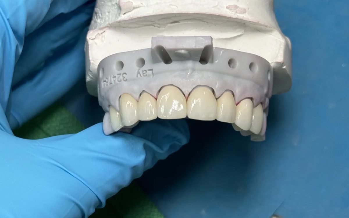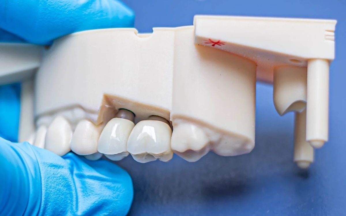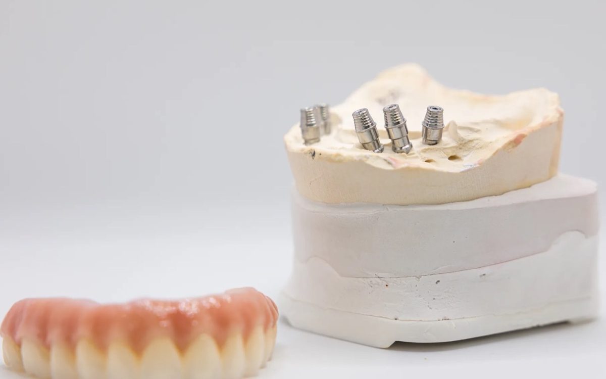
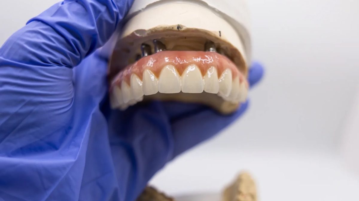
Photogrammetry dental capture records full arch implant positions with a camera system reading coded markers instead of stitching intraoral scan images together. On full arch cases that matters, because stitching error is exactly what makes long span intraoral scans unreliable. The 2025 evidence says photogrammetry is the more accurate of the two for implant position.
It does not say photogrammetry finishes the case on its own. Here is what it captures, what it does not, and how we would fit it into a full arch workflow from the lab side.
What is photogrammetry in dentistry?
Photogrammetry in dentistry is a method of recording the three dimensional position and angulation of multiple dental implants by photographing coded markers, often called scan flags, attached to each implant or abutment, and calculating their positions from the images. It replaces the implant position part of a full arch impression rather than the whole impression.
Because a photogrammetry camera measures every marker in one frame of reference, it avoids the image stitching that an intraoral scanner has to do across an edentulous arch. The output is a file of implant positions that the lab merges with a scan of the soft tissue and the opposing arch, which then becomes the basis for full arch zirconia bridges and hybrids. International Dental Arts builds full arch implant restorations for dentists nationwide, and our General Manager, Adam Brewer, has completed more than 2,500 full arch conversions with a focus on All-on-X work.
- What it records. Implant or abutment position and angulation, across the whole arch at once.
- What it does not record. Soft tissue, teeth, the bite and the vertical dimension.
- What it replaces. The splinted open tray impression or the long span scan of the implant positions.
- What it does not replace. The tissue scan, the bite records and the try-in.
Photogrammetry dental accuracy against intraoral scanning
The most useful single source is the 2025 systematic review and meta-analysis by Pozzi and colleagues in Clinical Implant Dentistry and Related Research. It included 13 studies, 3 in vivo and 10 in vitro, covering seven intraoral scanners and two photogrammetry systems, PIC and ICam4D, on cases with 4 to 8 implants.
Photogrammetry reported higher accuracy than intraoral scanning in 10 of the 13 studies. Meta-analyses of the in vitro studies found significant differences favouring photogrammetry in surface deviation trueness and precision and in angular deviation trueness (p below 0.05). Angular trueness ranged from 0.24 to 0.80 degrees with photogrammetry and from 0.28 to 1.74 degrees with intraoral scanning, and the authors noted that intraoral scanning produced angular deviation exceeding the 1 degree threshold associated with passive fit. The abstract is on PubMed.
Two cautions sit inside that result. Most of the evidence is in vitro, and the authors call for clinical trials before the question is settled. And their own closing recommendation is that a rigid prototype try-in is still recommended until then.
What photogrammetry does not solve
A photogrammetry dental record fixes one problem very well: where the implants are relative to each other. A full arch prosthesis also depends on things it never measures, and this is the stance we would hold on any full arch case: a perfect implant position file on top of a wrong bite is still a remake.
- Soft tissue form. The intaglio of a hybrid or a zirconia bridge is designed against the tissue scan, not the photogrammetry file.
- Vertical dimension and bite. These come from the records and the existing prosthesis, not from the implants.
- Tooth position and esthetics. Midline, smile line and lip support are decided at try-in.
- The merge. The photogrammetry file and the intraoral scan have to be aligned to each other, which is its own step with its own room for error.
That is why we read the 2025 recommendation for a prototype try-in as the right default rather than a formality. Photogrammetry makes the try-in more likely to be passive. It does not make it unnecessary.
You do not need photogrammetry to do full arch well
This is the second position worth stating plainly. It is easy to read the 2025 evidence as saying photogrammetry is the price of entry to full arch work. It is not. A splinted open tray impression in a rigid custom tray, or a verified intraoral scan, followed by a prototype try-in, remains a sound full arch workflow, and the verification step does the job of catching position error before the definitive prosthesis is made.
Photogrammetry is worth having if you do enough full arch cases for its accuracy and speed to pay back the cost of the system. If you do not, verification is the cheaper route to the same passive fit. Our scan body article covers the verification options in where full arch scanning runs out of road, and the abutment side in when you need a multi unit abutment.
Sending a photogrammetry case to the lab
A photogrammetry dental case arrives as several files that have to belong together. What we need is short, and most of it is the same as any full arch case.
- The photogrammetry export and the name of the system it came from.
- An intraoral scan of the soft tissue with the abutments or healing caps visible, and the opposing arch.
- The implant system and abutment level, usually multi-unit, with platform details.
- Bite and vertical records, or the existing prosthesis scanned in occlusion.
- What you want at try-in, a printed prototype or a verification step first.
If you are about to buy a photogrammetry system or start using one, tell us before the first case so we can confirm the file format and the merge workflow with you rather than discovering a mismatch when the files land.
Is a full arch case worth sending to a lab we haven’t used before?
Full arch is where a new lab relationship feels riskiest, so it is fair to ask. We have Clinical Specialists for All-on-X, implant, fixed and removable cases, and you can talk to one before the case is planned and while it is in production. That conversation is where the try-in plan, the abutment level and the material are decided, before anything is milled.
Our full arch zirconia work is on the zirconia bridges page linked above, and the wider implant range on implant restorations. When you are ready to send a case, start with us here.
Full arch implants
Planning a full arch case?
Talk to a Clinical Specialist before you capture. We will plan the records, the try-in and the material with you.
Questions from the prep
Is photogrammetry more accurate than an intraoral scan for full arch?
For implant position, the 2025 Pozzi meta-analysis says yes. Photogrammetry was more accurate in 10 of 13 studies, and intraoral scanning exceeded the 1 degree angular threshold associated with passive fit. Most of that evidence is in vitro.
Does photogrammetry replace the try-in?
No. The same review still recommends a rigid prototype try-in until clinical trials settle the question, and the try-in checks the bite, tissue fit and esthetics, which photogrammetry never measures.
Can photogrammetry be used for single implants?
It is designed for multiple implants across an arch, where stitching error is the problem it solves. A single implant or a short span is well served by a conventional impression or an intraoral scan, and photogrammetry adds cost without fixing a problem that case has.
What does the lab need besides the photogrammetry file?
A soft tissue scan, the opposing arch, bite and vertical records, and the implant system and abutment level. The photogrammetry file only tells us where the implants are. Everything that makes the prosthesis fit the patient, rather than the implants, comes from the other records, so a case with a perfect position file and no usable bite still cannot be built. If the patient is wearing an immediate or interim prosthesis that is working, scan it in the mouth and in occlusion as well. It carries the vertical dimension, tooth position and lip support the patient has already adapted to, and it is the fastest way for us to design a prototype that starts close to right instead of starting from nothing. Tell us which parts of it the patient likes and which they do not.
Written by Adam Brewer, General Manager, with nearly two decades in the dental implant industry and more than 2,500 full arch conversions, at International Dental Arts. IDA has been a family founded dental laboratory in Tulsa since 1984, working with independent practices across the country. If you want a case reviewed before you commit, get in touch.